Contents
Imaging Analysis of Brain Function
Since Descartes separated brain and mind in his theory of the dualism, the mind has been a subject of inquiry in philosophy, psychology, and religion. However, recent technical developments have shown that the mind, or at least some parts of it, can be explained materialistically. In line with that idea, we are making an earnest effort to further substantiate this hypothesis. We have adopted the following methodological approaches:
Imaging of Neurotransmission
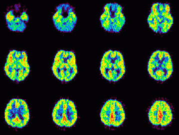
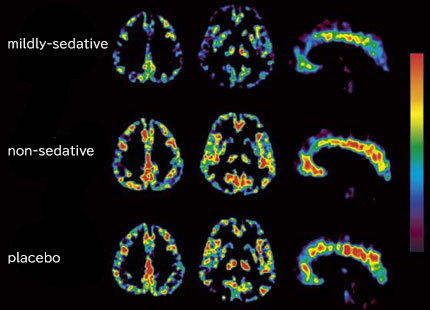
One of the advantages of PET over other brain imaging methods is its high sensitivity, which makes it suitable for neuronal receptor quantification. Previously, we mainly used [18F]fluoro-L-DOPA, [11C]YM9151-2, [11C]benztropine, and [11C]doxepin(Fig. 1)for imaging of the dopamine metabolism, D2 receptor, muscarinic acetylcholine receptor, and histamine H1 receptor distributions in human brain. We reported that dopamine D2 receptors are up-regulated in patients with vascular dementia. Clinical studies using [11C]doxepin is also being continued in order to evaluate the sedative side effects of various histamine H1 receptor antagonists (antihistamines). This series of studies has been accepted by the Consensus Guideline for New Generation Anthistamine (CONGA)(Fig. 2).
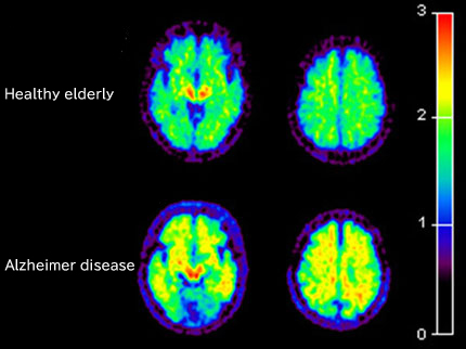
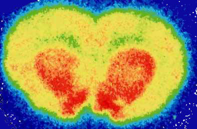
Furthermore, we put more enphasis on "molecular imaging" studies at CYRIC. Various new tracers have been evaluated for their clinical efficacies, such as [18F]FRP-170, for the imaging hypoxic cells, [11C]BF-227, for the imaging beta-amyloid deposition(Fig. 3), and [11C]donepezil, for the evaluation of function of acetylcholinergic nerves (Fig. 4).
Imaging of neuropsychological abnormalities in cancer patients
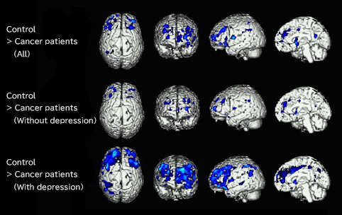
Whole body imaging of glucose metabolism has been routinely carried out for cancer diagnosis using 3D-PET. We analyzed regional brain metabolism in many patients having cancer and compared with age-matched control subjects. The SPM96 (Friston et al. J Cereb Blood Flow Metab. 11:690-699, 1991) was used for brain standardization and statistical comparisons. We have identified several regions with decreased FDG uptake in several brain regions like limbic and paralimbic areas. The results have suggested that PET is effective to objectively visualize psychological or emotional changes in the patients with severe ailment including cancer (Fig. 5).
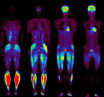
Imaging of energy redistribution during physical exercise
FDG is known to accumulate in accordance with regional energy consumption. When this tracer is applied during physical exercise, or before exercise, we can map and identify muscles of different compartments that participate in the action. We are convinced that PET imaging technique can assist sport science in designing optimum training programs. Moreover, this approach is useful in order to investigate the metabolism of all bodily organ, including brain. After analyzing the brain data with the SPM software, we identified brain regions that were activated during running. The primary visual and visual association cortices were the sites most actively involved during running (Fig. 6).